Home
/ Cross Section Of A Spongy Bone - Bone Structure | Anatomy and Physiology I / (these terms refer to their macroscopic appearance.)
Cross Section Of A Spongy Bone - Bone Structure | Anatomy and Physiology I / (these terms refer to their macroscopic appearance.)
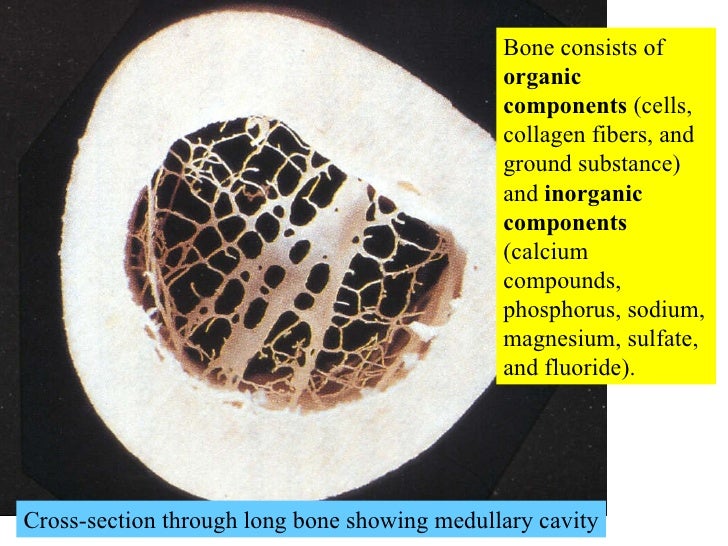
Cross Section Of A Spongy Bone - Bone Structure | Anatomy and Physiology I / (these terms refer to their macroscopic appearance.). It is highly vascularized and contains spongy bone is softer and weaker than compact bone, but is also more flexible. Structure of compact and spongy bone tissue. Bone that has many spaces in it, giving it a spongy appearance. Here is a section of decalcified spongy bone. Cross section of a human bone showing bone marrow, spongy bone and blood vessels.
Have you ever seen fossil remains of dinosaur and ancient human bones in cross section showing osteons. Anatomy of a thigh bone. (these terms refer to their macroscopic appearance.) Jump to navigation jump to search. Explaned distal and proximal epiphysis.
03 Cartilage And Bone Connective Tissue from image.slidesharecdn.com I also don't like how the bone and the marrow is. For example, in the human cancellous bone in which the cross struts are disconnected (c) is not very capable of supporting a. Osteoclasts or bone resorbing cells. Here is a section of decalcified spongy bone. (these terms refer to their macroscopic appearance.) Two types of bone tissues in cross section of a long bone : As the names suggest compact bone looks compact and the spongy bone looks like sponges. Transverse cross section of compact bone tissue;
Spongy bone has many spaces filled with red bone marrow that provide the osteocytes with nutrients.
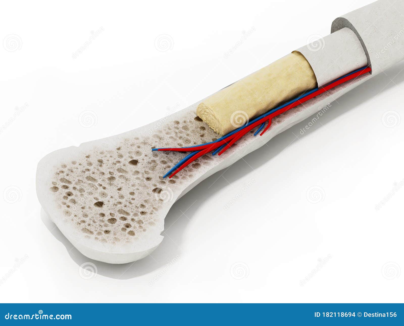
Osteoclasts or bone resorbing cells. Cross section of a human bone showing bone marrow, spongy bone and blood vessels. Blue arrows indicate lacunae spongy bone, also known as cancellous bone or trabecular bone, looks like a sponge under the microscope. Internal structure of a human long bone, with a magnified cross section of the interior. Note while here the possible presence of osteoblasts and osteoclasts in spongy bone. The large dark spots are passages for blood vessels and nerves. For example, in the human cancellous bone in which the cross struts are disconnected (c) is not very capable of supporting a. Two types of bone tissues in cross section of a long bone : Diagram with articular cartilage, marrow, spongy bone, medullary cavity, endosteum, diaphysis, and. Transverse cross section of compact bone tissue; Frontal section, anterior and posterior view. Tubular bone and spongy bone structure. Spongy bone also contains osteocytes housed in lacunae.
Jump to navigation jump to search. Osteoblasts and osteocytes, responsible for creating bone. Blue arrows indicate lacunae spongy bone, also known as cancellous bone or trabecular bone, looks like a sponge under the microscope. Osteoporosis causes a reduction in the amount of bone mass, including rarefaction of spongy bone tissue and cortical erosion here, of a lumbar. Internal structure of a human long bone, with a magnified cross section of the interior.
Bone cross-section, SEM - Stock Image - C019/5246 ... from media.sciencephoto.com Osteoclasts or bone resorbing cells. Spongy bone is the inner framework of the bone in which the bone marrow resides. The red arrow indicates a haversian canal; When we look at the blood pathways, what we're looking at is. Because this bone is not dense, spongy bone needs the protection of a thin, outer layer of compact bone. Explaned distal and proximal epiphysis. The compact bone is made up of osteon. A cross section of a human long bone.
The canaliculi connect to the adjacent cavities, instead of a central haversian canal, to receive their.
Note while here the possible presence of osteoblasts and osteoclasts in spongy bone. Diagram with articular cartilage, marrow, spongy bone, medullary cavity, endosteum, diaphysis, and. The canaliculi connect to the adjacent cavities, instead of a central haversian canal, to receive their. Cancellous bone , also called trabecular bone or spongy bone , light, porous bone enclosing numerous large spaces that give a honeycombed or spongy appearance. Tubular bone and spongy bone structure. Spongy (cancellous) bone is lighter and less dense than compact bone. The mineral that is found in the extracellular. The red arrow indicates a haversian canal; Jump to navigation jump to search. Spongy bone is the inner framework of the bone in which the bone marrow resides. Long bone, compact bone and spongy bone. Anatomy of a thigh bone. Spongy bone consists of plates (trabeculae) and bars of bone adjacent to small, irregular cavities that contain red bone marrow.
Here is a section of decalcified spongy bone. The red arrow indicates a haversian canal; Diagram with articular cartilage, marrow, spongy bone, medullary cavity, endosteum, diaphysis, and. Blue arrows indicate lacunae spongy bone, also known as cancellous bone or trabecular bone, looks like a sponge under the microscope. (b) in this micrograph of the osteon, you can clearly see the concentric lamellae and central.
Cross Section Of A Human Bone Showing Bone Marrow, Spongy ... from thumbs.dreamstime.com It is highly vascularized and contains spongy bone is softer and weaker than compact bone, but is also more flexible. At this stage of development, they are most likely to be along the outermost (under the periosteum) or innermost (next to the bone marrow) surfaces of the bone; It is lighter, less dense, and more flexible than compact bone. A cross section of a human long bone. Have you ever seen fossil remains of dinosaur and ancient human bones in cross section showing osteons. Spongy bone also contains osteocytes housed in lacunae. Long bone, compact bone and spongy bone. Spongy bone has many spaces filled with red bone marrow that provide the osteocytes with nutrients.
Cancellous (trabecular or spongy) bone: Have you ever seen fossil remains of dinosaur and ancient human bones in cross section showing osteons. Cancellous bone , also called trabecular bone or spongy bone , light, porous bone enclosing numerous large spaces that give a honeycombed or spongy appearance. Frontal section, anterior and posterior view. Two types of bone tissues in cross section of a long bone : □ compact tissue, it is dense in texture and it is always placed on the □ the osteon consists of a system of bony lamellae arranged concentrically around a canal, which is called haversian canal and this canal. Here is a section of decalcified spongy bone. Because this bone is not dense, spongy bone needs the protection of a thin, outer layer of compact bone. Internal structure of a human long bone, with a magnified cross section of the interior. Spongy (cancellous) bone is lighter and less dense than compact bone. □ on examining a cross section of any bone, it is composed of two kinds of bony tissue: Bone that has many spaces in it, giving it a spongy appearance. There are trabeculae in spongy bone which gives its sponge like appearance.
Osteoblasts and osteocytes, responsible for creating bone cross section of a bone. It is lighter, less dense, and more flexible than compact bone.